
Figure 7.
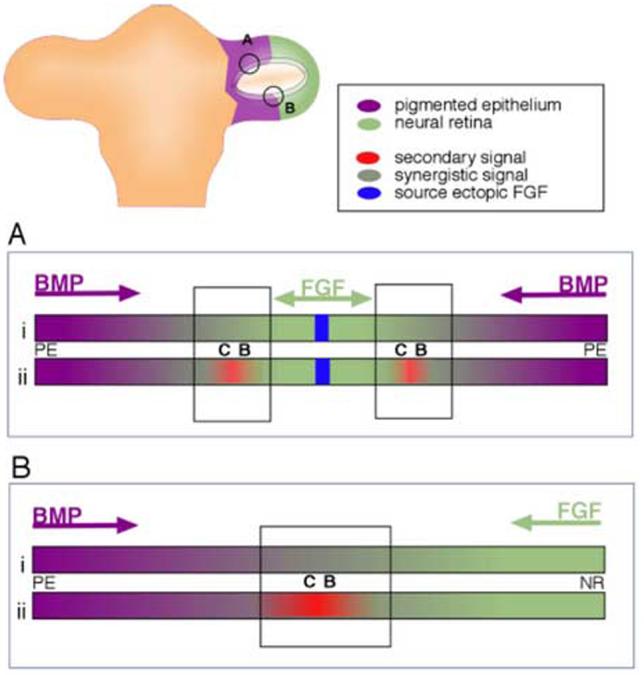
Model for establishment of ciliary body tissue during optic vesicle stages. (A) Ciliary body tissue is created at the edges of ectopic patches of neural retina, as demonstrated in this study. Hypothetically, this might occur either directly (i) through overlapping BMP and FGF signals, or indirectly (ii), through the creation of a second signaling center (red), that itself is created through overlapping BMP and FGF signals. (B) Extrapolation of experimental result to the establishment of the ciliary body at the border between the neural retina and the pigmented epithelial domains in the optic vesicle.