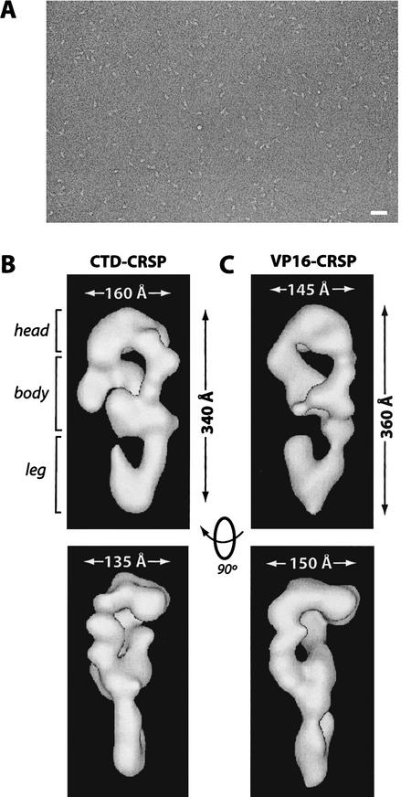
Figure 3.
CTD–CRSP and VP16–CRSP are structurally similar. (A) Negatively stained electron micrograph of CTD–CRSP sample. Bar, 800 Å. (B,C) Three-dimensional reconstruction of CTD–CRSP and VP16–CRSP at 32 Å resolution. Complexes are rendered to 1.25 MD, their approximate predicted molecular weight. Dimensions shown. Rotation of the volumes 90° gives the second side view of the coactivator.

