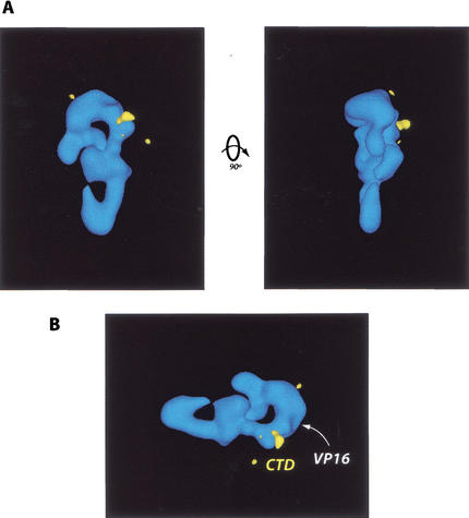
Figure 4.
(A) Localization of the CTD binding site (yellow) on the CRSP coactivator. This site was identified via EM analysis and difference mapping of structures generated from CTD–CRSP samples incubated with anti-GST antibodies, which target the GST–CTD fusion protein bound to the CRSP complex. (B) VP16 and CTD bind similar regions on the CRSP complex. As in A, the CTD-binding site is shown in yellow. The VP16-binding site is indicated by the white arrowhead.

