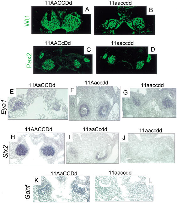
Figure 5.
Molecular characterization of Hox11 triple mutant phenotype. (A–L) E10.5 frontal sections through the metanephric region. (A,B) Immunohistochemical staining of Wt1 in control and triple mutant embryos in the metanephric blastema and surrounding urogenital mesenchyme. Note that the control section in A is at a slightly later stage, approximately E11. At the left side of A, the ureter (which does not stain for Wt1) can be seen entering the metanephric mesenchyme. (C,D) Pax2 immunostaining in the blastema and Wolffian duct. There are no observable differences between control and mutant embryos. (E–G) Eya1 expression as detected by in situ hybridization (ISH). (E) Control embryo. (F) Five-allele mutant, 11Aaccdd, shown at a slightly later stage (approximately E11, note invasion of both blastemas by nonexpressing ureteric bud). (G) Triple homozygous mutant. Expression of Eya1 is normal in mutants and controls. (I) Six2 ISH demonstrates a drastic reduction in Six2 message in five-allele embryos (11aaCcdd shown here at approximately E11, note invasion of the right metanephric blastema by nonexpressing ureteric bud), compared to control expression (H). There is a complete loss of message in triple mutant mesenchyme (J) compared to control (H). Gdnf expression as detected by ISH in control (K) and triple mutant (L) is absent or much reduced in triple mutant blastemas.

