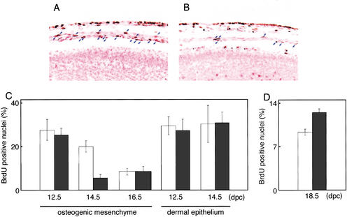
Figure 5.
Reduced proliferation of osteogenic cells and increased proliferation of chondrocytes in Fgf18−/− embryos. (A,B) BrdU labeling in the mediodorsal regions of the crania of wild-type (A) and Fgf18−/− (B) embryos in the same litter at 14.5 dpc. Blue arrows indicate osteogenic mesenchymal cells labeled with BrdU. Counterstaining was performed with nuclear fast red. Artifactual separation of the layers of osteogenic mesenchyme from the dermal epithelium and neuroepithelium occurred during the process of fixation. (C,D) The proportion of BrdU-positive cells in osteogenic mesenchyme and in dermal epithelium in the mediodorsal regions of the crania at 12.5, 14.5, and 16.5 dpc (C) and in the proliferating chondrocytes at 18.5 dpc (D) of wild-type (open bars) and Fgf18−/− (solid bars) mice.

