Fig. 1.
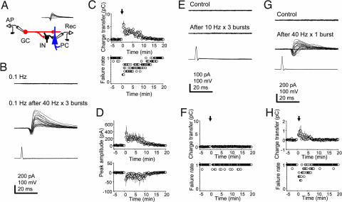
Granule cell (GC) bursting results in short-term facilitation of synaptic transmission. (A) Experimental configuration for recording from a GC–pyramidal cell (PC) pair in a mossy-fiber unitary circuit. (Inset) Biphasic mossy-fiber responses evoked in a targeted CA3 PC (−70 mV) by single APs in a GC at 1 Hz. (B) Single APs induced at 0.1 Hz in a GC (Bottom) fail to evoke responses in a CA3 PC (Top). After 40-Hz bursts of APs in the GC (three trains of 2.5-s duration every 10 s), single APs at 0.1 Hz reliably evoke unitary responses in the PC for minutes (Middle). (C) (Upper) Plot of the evoked responses (calculated charge transfer) over time (n = 7). The arrow indicates the time when GC bursting was induced. (Bottom) Plot of pooled synaptic failures over time (n = 7). The seemingly quantal jumps in the failures plot reflect the pooled all-or-none responses from the individual cells. (D) Peak amplitude of currents over time is shown for the IPSCs and the EPSCs, respectively. (E) Single APs induced at 0.1 Hz in a GC fail to evoke responses in a synaptically connected CA3 PC, both before (Top) and after (Middle) 10-Hz bursts of APs in the GC (three trains, 10-s duration). (F) Plots of charge transfer and failure rates over time. (G) Single APs induced at 0.1 Hz in a GC that fail to evoke responses in a synaptically connected CA3 PC successfully evoke responses after a single 40-Hz burst of 2.5 s in the GC. (H) Plots of charge transfer and failure rates over time. [Scale bars: vertical, 200 pA (PC, B and G), 100 pA (PC, E), 100 mV (GC); horizontal, 20 ms.]

