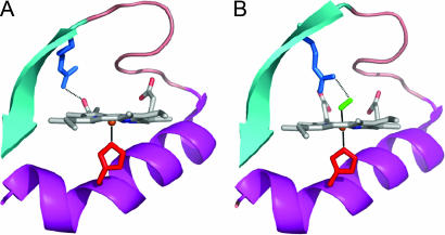
Fig. 1.
Crystallographic structures of the ferrous deoxy complex [Protein Data Bank (PDB) ID no. 1LSW; ref. 4] (A) and oxycomplex (PDB ID no. 1DP6; ref. 3) (B) of FixLH. The residues shown are the proximal histidine (H200, red) and the distal argine (R220, blue). The latter forms a salt bridge with heme propionate 7 in the deoxy form and a hydrogen bond with O2 (green) in the oxy complex. The figure was prepared using PYMOL.

