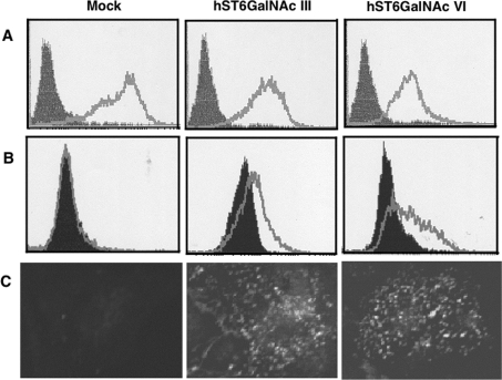
Figure 3. Expression of DSGG in Caki-1 cells transfected with hST6GalNAc III and VI cDNA.
(A and B) Flow cytometric patterns of Caki-1 cells transiently transfected with empty pcDNA3.1 vector (Mock), pcDNA3.1-hST6GalNAc III or VI. The expression of MSGG (A) and DSGG (B) in Caki-1 cells was detected with mAbs RM1 and 5F3 at a 1:1 dilution (cell culture supernatant) respectively. (C) Microscopic fluorescence images of Caki-1 cells transiently transfected with empty pcDNA3.1 vector, pcDNA3.1-hST6GalNAc III or VI. The cells were fixed with paraformaldehyde and permeabilized with 0.1% Triton X-100 and then processed for indirect immunofluorescence analysis as described in the Materials and methods section. Original images were obtained at 400× magnification.

