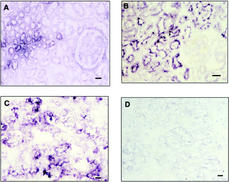
Figure 7. Imunohistochemical staining of normal human kidney and renal cancer tissues from the same cases.
(A) Normal kidney section stained by mAb RM1 (100× magnification). (B) Normal kidney section stained by mAb 5F3 (200× magnification). (C) Cancer section stained by mAb RM1 (100× magnification). (D) Cancer section stained by mAb 5F3 (100× magnification). Scale bar: 40 μm.

