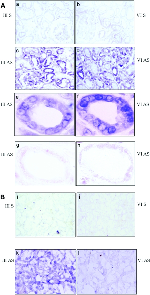
Figure 9. In situ hybridization using PNA probe for hST6GalNAc III/VI in non-tumour kidney and tumour sections.
(A) Non-tumour kidney sections. Panel a, sense probe of hST6GalNAc III. Panel b, sense probe of hST6GalNAc VI. Panel c, anti-sense probe of hST6GalNAc III. Panel d, anti-sense probe of hST6GalNAc VI. Panels e and f, the proximal tubule shown by higher magnification of panels c and d respectively. Panels g and h, the distal tubule shown by higher magnification of c and d respectively. (B) Tumour sections. Panel i, sense probe of hST6GalNAc III. Panel j, sense probe of hST6GalNAc VI. Panel k, anti-sense probe of hST6GalNAc III. Panel l, anti-sense probe of hST6GalNAc VI. Original magnifications are as follows, panels a–d, 200×; panels e–h, 1,000×; and panels i–l, 200×. S, sense; AS, antisense.

