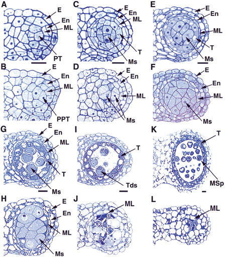
Figure 2.
Comparison of the wild-type and ems1 mutant anther development. The panels show the upper right lobe of the anther. (A) Wild-type anther at stage 4 with the epidermis, endothecium, middle layer, and precursors of tapetal cells; cells of a particular layer are not formed simultaneously. (B) Stage-4 mutant anther with a structure similar to the wild-type in A. (C) Wild-type anther at early stage 5 showing the epidermis, endothecium, middle layer, tapetal layer, and microsporocytes. (D) Mutant anther at early stage 5 lacking the tapetal layer. (E) Wild-type anther at late stage 5 with structure similar to that in C. (F) Mutant anther at late stage 5 with excess microsporocytes and a more obvious absence of the tapetal layer. (G) Stage-6 wild-type anther with epidermis, endothecium, degenerated middle layer, vacuolated tapetal cells, and isolated microsporocytes. (H) Mutant anther at stage 6 containing enlarged and undetached microsporocytes. The middle layer had not degenerated. (I) Stage-7 wild-type anther showing tapetal layer and tetrads. (J) Stage-7 mutant anther with degenerating microsporocytes and lacking tapetal layer. The middle layer was still present. (K) Wild-type anther at stage 9 with tapetal layer and microspores. (L) Stage-9 mutant anther showing degenerated microsporocytes and persistent middle layer. E, epidermis; En, endothecium; ML, middle layer; Ms, microsporocytes; MSp, microspores; PT, precursors of tapetal cells; PPT, putative precursors of tapetal cells; T, tapetal layer; Tds, tetrads. Bar, 10 μm. A and B, C and D, E and F, G and H, I and J, K and L have same magnification.

