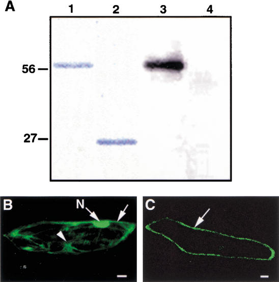
Figure 5.
Kinase activity analysis of the EMS1 protein and subcellular localization of an EMS1-GFP fusion protein. (A) Autophosphorylation activity of the EMS1 kinase domain. (Lane 1) A fusion protein between the EMS1 kinase domain and GST. (Lane 2) GST protein. (Lane 3) The fusion protein between the EMS1 kinase domain and GST showing kinase activity of autophosphorylation. (Lane 4) GST alone having no activity of autophosphorylation. (B) A cell that expressed free GFP showing fluorescence in nucleus (N), cytoplasm (arrowhead), and plasma membrane (arrow). (C) A cell that expressed EMS1-GFP showing fluorescence in the plasma membrane (arrow). Bar, 25 μm.

