
Figure 6.
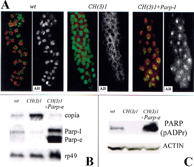
Expression of Parp-I or Parp-e cDNA rescues defects in CH(3)1 mutants. (A) Partial restoration of normal nuclear morphology by expression of Parp-I. Immunofluorescent detection of the nucleolar antigen AJ1 (red) and DNA (green) is shown in larval salivary glands of the indicated genotypes. AJ1 staining alone is shown on the right. In CH(3)1 mutants (middle), AJ1 is cytoplasmic rather than in nucleoli as in wild-type (left). Expression of Parp-I cDNA (right) restores nucleoli and nuclear AJ1 staining in a mosaic manner; note cells at the top of the figure with normal localization, but cells near the bottom still show a mostly cytoplasmic distribution of AJ1 reactivity. (B) A Northern blot of RNA from larvae of the indicated genotypes shows that Parp-e cDNA expression greatly elevates the level of 2.6-kb Parp-e mRNA and also of the 3.2-kb Parp-I mRNA. Note that copia-specific RNA accumulation is greatly reduced in CH(3)1 mutant larvae that express Parp-e cDNA. rp49 hybridization serves as a loading control. (C) A Western blot of proteins isolated from larvae of the same genotypes as in C, and probed with an antibody specific for poly(ADP-ribosyl) moieties. Expression of Parp-e cDNA in a CH(3)1 homozygous background increases the amount of poly(ADP)-ribose-modified proteins to levels greater than in wild-type. As in wild-type, diverse proteins are affected, the most prominent of which is the size of PARP-I itself (shown). An actin antibody was used as a loading control.