
Figure 7.
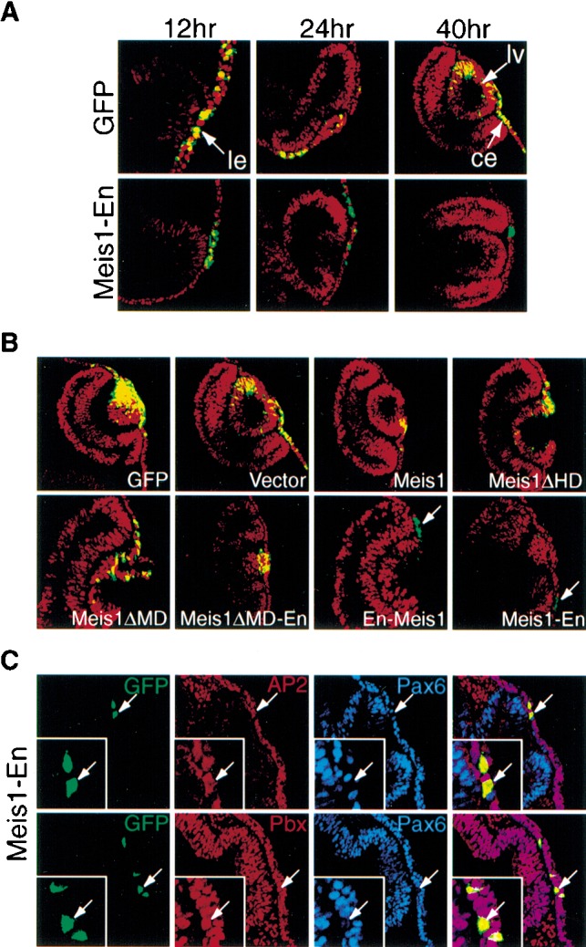
Meis1-En represses Pax6 lens ectoderm expression in vivo. (A, top) In chick embryos, endogenous Pax6 expression (red fluorescence) is not altered by electroporation of a Control GFP-expressing vector (green fluorescence), as evident by extensive overlapping expression (yellow). (Bottom) In contrast, following co-electroporation of Meis1-En (10-fold molar excess of a construct expressing Meis1 fused to the Drosophila Engrailed repressor domain) and GFP, endogenous Pax6 nuclear expression in the prospective lens ectoderm is reduced by 12 h and 24 h, and Pax6−/GFP+ cells are excluded from the lens vesicle by 40 h. (ce) Corneal ectoderm; (le) lens ectoderm; (lv) lens vesicle. (B) Requirement of the full-length Meis1-Engrailed repressor domain protein to repress Pax6 expression. When chicken embryos were electroporated with GFP plus various Meis contructs (the latter present in 10-fold molar excess) and analyzed after 24 h, only En-Meis1 and Meis1-En repressed Pax6 expression. Pax6+ cells are red, Meis construct expressing cells are green (due to GFP), and Pax6+/GFP+ cells are yellow. (C) Meis1-Engrailed proteins do not repress AP2α or Pbx1–3 expression. After electroporation, Meis1-Engrailed positive cells (GFP+, green) still express AP2α (red) and Pbx (red), but not Pax6 (blue).