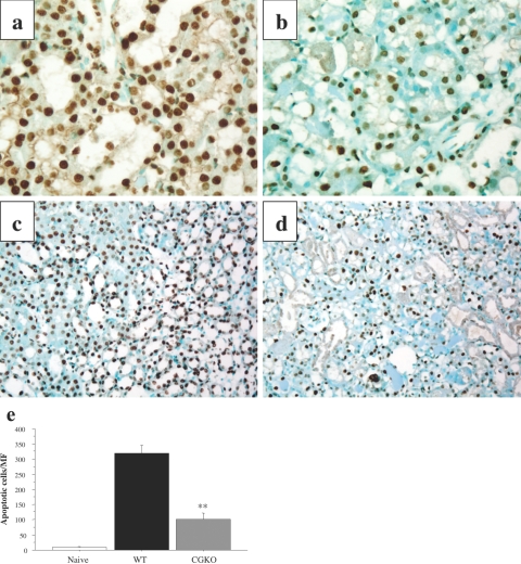
Figure 8.
Decreased apoptosis in kidneys subjected to I/R injury in the absence of cathepsin G. Wild-type 129/SvJ (a and c) and cathepsin G−/− (b and d) mice were subjected to 60 minutes of warm renal ischemia. After 24 hours of reperfusion, the ischemic kidneys were removed, and formalin-fixed sections were prepared and stained for TUNEL labeling to detect apoptotic cells. Magnification: ×400 (a and b); ×100 (c and d). e: The numbers of TUNEL-positive cells were counted in 10 microscopic (magnification, ×200) fields/slide for four slides/kidney and four kidneys per group. The mean positive number per field ± SD is shown for each group. **P < 0.001.

