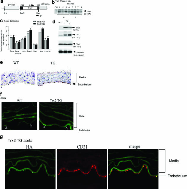
Figure 1.
EC-specific expression of Trx2. a: Schematic diagram for EC-specific transgenic construct in which expression of the HA-tagged Trx2 transgene is driven by a promoter sequence from the VE-cadherin gene. The native mitochondrial signal peptide (msp) on Trx2, and the primers for genotyping are indicated. b: Trx2 TG founders. Tails from the Trx2 transgenic founders (four males and four females based on genotyping with specific primers) were further analyzed for Trx2 expression by Western blot with anti-HA antibody. A nontransgenic tail was used as a control. c: Tissue distribution of the Trx2 transgene. Expression of the Trx2 transgene in different tissues was determined by a real-time RT-PCR using primers recognizing both human and murine Trx2. 18S rRNA was used for normalization. Data presented are fold increase in Trx2 mRNA in Trx2 TG lines (lines 1 and 3) compared with WT mice (n = 3). d: Expression of Trx2 protein in aorta. Trx2 protein in aorta was determined by Western blot with anti-HA or anti-Trx2 antibody. Trx1 was also detected by Western blot with anti-Trx1 antibody as a control. β-Tubulin was used as protein loading control. e--g: Trx2 transgene is expressed in the aortic endothelium. The Trx2 transgene in WT and Trx2 TG (line 3) aortic sections were detected by immunohistochemistry with anti-HA antibody. e: Endothelium and media are indicated. f: The Trx2 transgene in aortic sections were detected by indirect immunofluorescence microscopy with anti-HA antibody followed by Alexa Fluor 488 (green)-conjugated anti-mouse secondary antibody. Trx2 TG aorta were co-stained with anti-HA antibody followed by Alexa Fluor 488 (green)-conjugated anti-mouse secondary antibody and anti-CD31 antibody followed by Alexa Fluor 594 (red)-conjugated anti-goat secondary antibody. g: The merged picture is shown on the right.

