Figure 2.
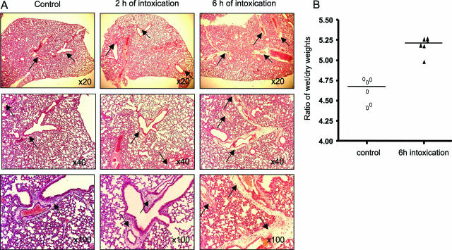
Histopathological analysis of the lung of TcsL-82-treated Swiss mice. A: H&E staining of lungs from HBSS-injected control mice and from mice intoxicated for 2 and 6 hours with intraperitoneally injected TcsL-82 (15 ng/mouse). Lungs were examined with ×20, ×40, and ×100 lenses. Perivascular space corresponding to the area where exudate is observed after intoxication (×20 lens and higher magnifications) is very limited in control lung, increased already after 2 hours, and is massive after 6 hours of intoxication (arrows). B: Ratios of wet-to-dry weights of lung from control and intoxicated mice with TcsL-82 (15 ng/mouse, i.p.). Horizontal bars indicate the median values. The values of control versus intoxicated mice were analyzed by the Wilcoxon signed-rank test using arbitrarily the median of the control population as the hypothetical value for comparison; P = 0.0156.

