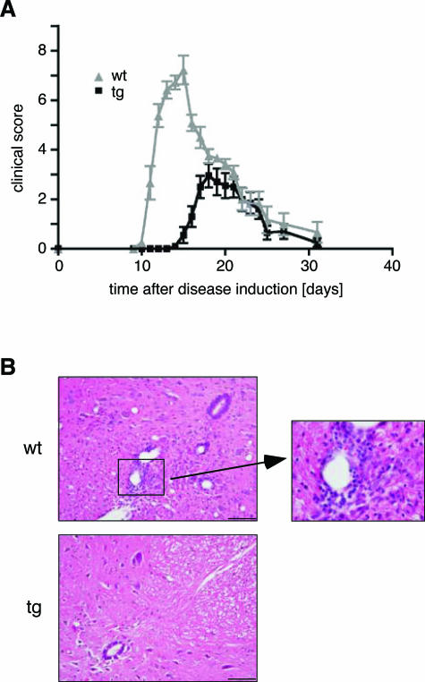
Figure 6.
Experimental autoimmune encephalomyelitis. A: Clinical disease scores for 3-month-old transgenic rats (black line, N = 10) and wild-type littermates (gray line, N = 9). One representative experiment of four is shown. B: H&E staining of paraffin sections from the lumbar spinal cord obtained at day 13 after EAE induction (N = 3). Scale bar = 100 μm. A highly infiltrated part of the wild-type spinal cord is enlarged.

