Figure 6.
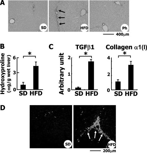
Induction of liver fibrosis in HFD-fed rabbits. A: Sirius Red staining of liver sections. Note that collagen fibers extended from the portal vein area in HFD-fed rabbits. Ph, HFD-fed rabbits treated with phlebotomy. B: Hydroxyproline content of the liver. To estimate the total amount of collagen in the liver, the level of hydroxyproline was measured as described in Materials and Methods. *P < 0.01. C: Hepatic expression of TGF-β1 and collagen α1(I) mRNAs was determined by quantitative RT-PCR. D: Detection of SM α-actin by immunohistochemistry. In the liver of SD-fed rabbits, SM α-actin was faintly detected along the sinusoids and around portal areas. In the HFD-fed group, its expression was greatly augmented along the fibrotic septum extending from the portal area. Scale bars: 400 μm (A); 200 μm (D).

