Figure 7.
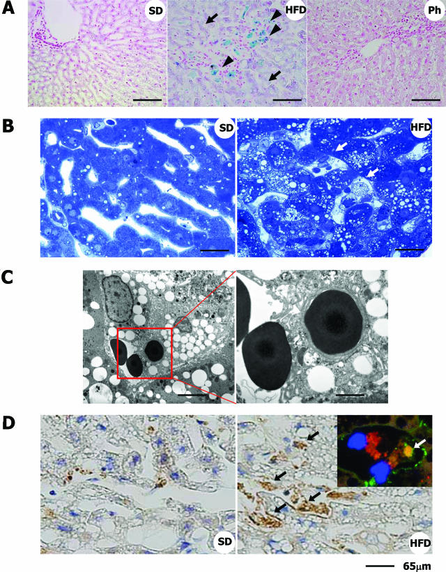
Iron deposition and increased erythrophagocytosis in the liver of HFD-fed rabbits. A: Berlin Blue staining. Note that iron is accumulated predominantly in macrophages (blue coloration, arrowheads) and in hepatocytes (purple coloration, arrows). Ph, HFD-fed rabbits treated with phlebotomy. (B) Toluidine blue staining of the liver. Note that macrophages in the sinusoids show foamy degeneration and are attached by erythrocytes (arrows). C: Electron microscopy. An erythrocyte is attached to a foamy macrophage, and another is in the phagosome of the macrophage. D: Detection of hemoglobin-α by immunohistochemistry. In normal liver, a faint scattering of hemoglobin-α was seen. In the liver of HFD-fed rabbits, its expression was greatly augmented in the Kupffer cells (black arrows). Inset shows the localization of hemoglobin-α (green, white arrow) in RAM-11-positive macrophages (red) in HFD-fed rabbit liver sections determined using fluorescence microscopy. Blue color indicates nucleus (DAPI staining). Scale bars: 100 μm (A); 40 μm (B); 10 μm (C, left); 3.5 μm (C, right); 65 μm (D).

