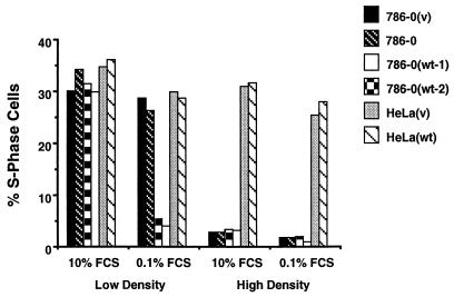
Figure 2.
Effect of cell density on growth. Two clones of 786–0(wt), one 786–0(v), and the parental 786–0 cell line as well as HeLa(v) and HeLa(wt) cell lines were plated at low (2–3 × 10⁵ cells per 15-cm dish) or high (2–3 × 10⁶ cells per 15-cm dish) cell density in the presence of 10% serum for 36 h. The cells were then either incubated in 10% serum (changing the serum every 12 h for 48 h) or switched to 0.1% serum for 48 h. Percentages of S phase cells were assessed by flow cytometry such as in A.

