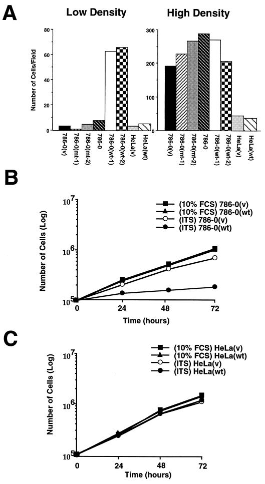
Figure 3.
Effect of cell density on cell death induced by the absence of serum. (A) Cells were plated to low or high density in 10% serum for 16 h followed by a 48- to 72-h incubation in DMEM/0% serum. Cells were then incubated for 10 min with Hoechst #33325, which stains nuclei of living cells. The numbers of live cells were determined by counting round, nonpycnotic nuclei per field. Experiments were performed with two wild-type clones, one 786–0(v), two 786–0(mt), and the parental 786–0 cell lines as well as HeLa(v) and HeLa(wt) cell lines. (B) Effect of ITS on the growth of 786–0(v) and 786–0(wt) cells in serum free media. Cells were plated at low density in DMEM/10% serum for 16 h followed by an incubation in either DMEM/10%FCS or DMEM/ITS. (C) Growth curves of HeLa(v) and HeLa(wt) cell lines incubated in either DMEM/10%FCS or DMEM/ITS. Cells were washed several times with DMEM, detached by trypsinization, and counted with a hemocytometer. Number of cells per 10-cm Petri dish was counted at the indicated time. The average of two independent experiments is shown.

