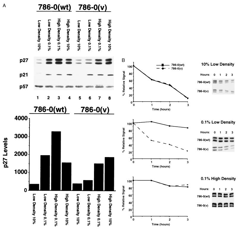
Figure 4.
Assessment of CKI levels and stability in 786–0(v) and 786–0(wt) cells. (A) 786–0(wt) and 786–0(v) cells were plated either at low or high cell density in 10% serum and incubated for 16–24 h followed by an incubation for 48 h in 0.1% or 10% serum. p27, p21 and p57 levels were analyzed by immunoblot analysis, and p27 levels were quantitated by densitometry of different exposures of autoradiographs. Immunoblotting with several anti-p27 antibodies revealed a p27 doublet. Both of these bands behaved identically in all experiments. (B) 786–0(wt) and 786–0(v) cells were plated in 10% serum and incubated for 48 h at low density in 10% FCS (Top), low density in 0.1% FCS (Middle), or high density in 0.1% FCS (Bottom). p27 levels were analyzed by immunoblot analysis. p27 stability was assessed by incubation of the cells in 100 μg/ml of cycloheximide for the indicated time periods. p27 levels were quantitated by densitometry of different exposures of autoradiographs.

