
FIG. 1. SNAP-29-EGFP is present in the synapses of cultured hippocampal neurons.
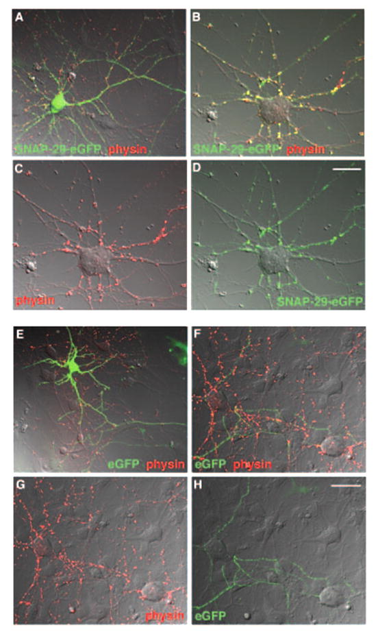
Cultured hippocampal neurons at DIV 7 were transfected with SNAP-29-EGFP (green, A–D) or EGFP (green, E–H) and immunostained with antibody against synaptophysin (physin, red), as a marker of synapses, after 24–48 h of transfection. Images are shown as merged differential interference contrast. A and E, transfected neurons. B–D and F–H, untransfected neurons that form synapses with transfected neurons. Note that presynaptically expressed SNAP-29-EGFP was targeted to the synapses with the untransfected neuron (B) and that EGFP is not significantly co-localized with synaptophysin at synapses (F). Scale bar, 20 μm.