
FIG. 3. SNAP-29 has no effect on the density of synapses in cultured hippocampal neurons.
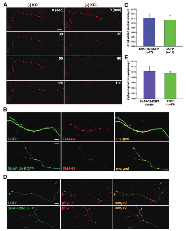
A, high K+-dependent FM4-64 destaining along axonal processes. Axon terminals loaded with FM4-64 were destained in the absence (left column) or presence (right column) of 90 mM KCl. The fluorescence images of FM4-64 were taken at different time points (0, 30, 60, and 120 s). B, axon terminals of hippocampal neurons at DIV 9–10 were loaded with FM4-64 (red) 24–48 h after transfection with SNAP-29-EGFP or EGFP (green). C, quantification of the relative density of FM dye-loaded synapses in neurons transfected with SNAP-29-EGFP (0.12 ± 0.018/μm) or EGFP control (0.11 ± 0.019/μm, mean ±S.E.; p > 0.05). D, immunostaining of the synaptic vesicle marker synaptophysin (physin, red) in neurons expressing SNAP-29-EGFP or EGFP (green) was used to determine the number of synapses. Cultured neurons at DIV 9–10 were obtained 24–48 h after transfection. E, quantification of the relative density of synaptophysin staining puncta in neurons transfected with SNAP-29-EGFP (0.10 ± 0.020/μm) or EGFP (0.096 ± 0.0062/μm; p > 0.05).