
Figure 5.
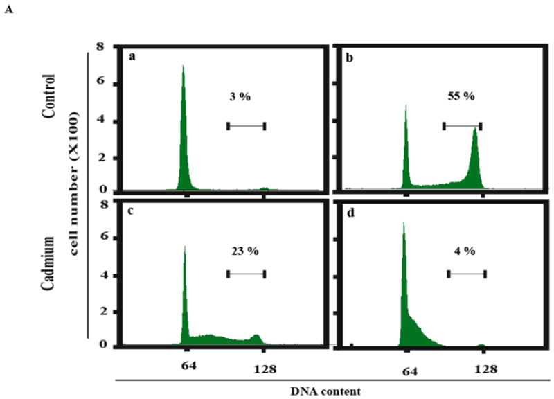
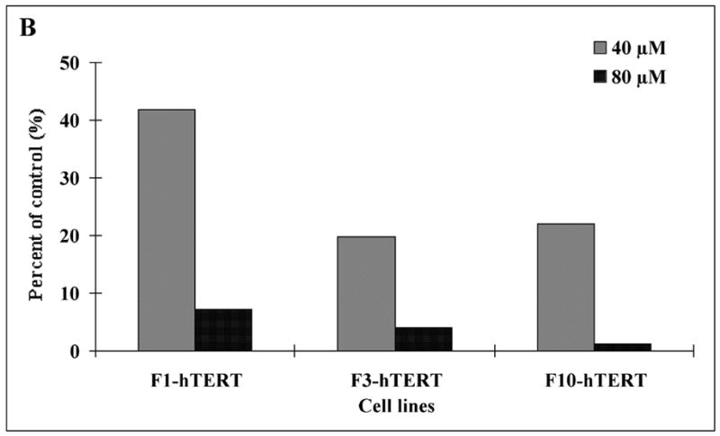
Inhibition of DNA synthesis in S phase cells. Cells were released from confluence-arrest, reseeded at 1 million cells per 100 mm dish and incubated with aphidicolin for 24 h to collect cells at the beginning of S phase. Aphidicolin was then removed and cells incubated in fresh medium with or without cadmium for 6 h to observe the increment of DNA content during S phase progression. Propidium iodide was used to stain DNA. The stained nuclei were analyzed by flow cytometry to determine their DNA content. (A) F1-hTERT cells were released from synchronization and treated with various concentrations of cadmium. Numbers above the bar (3–4 N DNA content) represent the fraction of total cells in this region. (a) Cells synchronized for 24 h with aphidicolin. (b) Cells incubated for 6 h without cadmium after removal of aphidicolin. (c) Cells incubated 6 h with 40 μM cadmium after removal of aphidicolin. (d) Cells incubated 6 h with 80 μM cadmium after removal of aphidicolin. (B) The percentage of nuclei with 3–4 N DNA content after 6 h incubation with 40 or 80 μM cadmium was expressed as a percentage of the sham-treated control.