
Figure 4. Thymocytes exposed to JWH-015 in vitro undergo apoptosis.
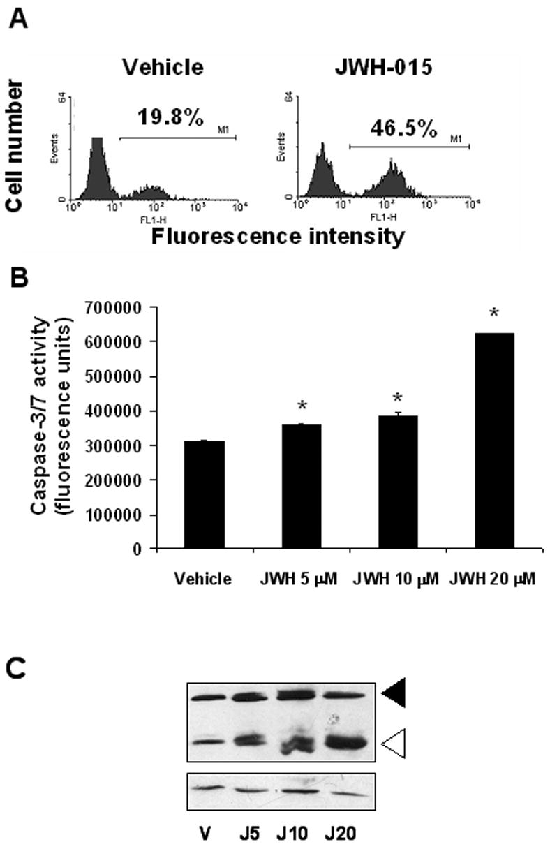
A) Thymocytes (5x10⁶) from C57BL/6 mice were incubated with 20 μM JWH-015 or the vehicle for 24hrs. The cells were harvested and analyzed for apoptosis using the TUNEL assay followed by flow cytometric analysis. The percentage of apoptotic cells was depicted on each histogram. B) Thymocytes from C57BL/6 mice were incubated with various concentrations of JWH-015 (5, 10, and 20 μM) or the vehicle overnight. The induction of apoptosis was then analyzed using the caspase-3/7 Apo-One fluorimetric assay. The data represent the mean ± SEM of duplicate cultures. Asterisk indicates statistically significant difference when compared to vehicle control. C) Thymocytes from C57BL/6 mice were cultured with various concentrations of JWH-015 (5, 10, or 20 μM) or the vehicle for 18hrs, after which, whole proteins were extracted and analyzed for caspase-3 cleavage by Western blotting. Beta-actin was used as a loading control.