
Figure 6. Gastric expression of exogenous FGF10 mRNA and protein.
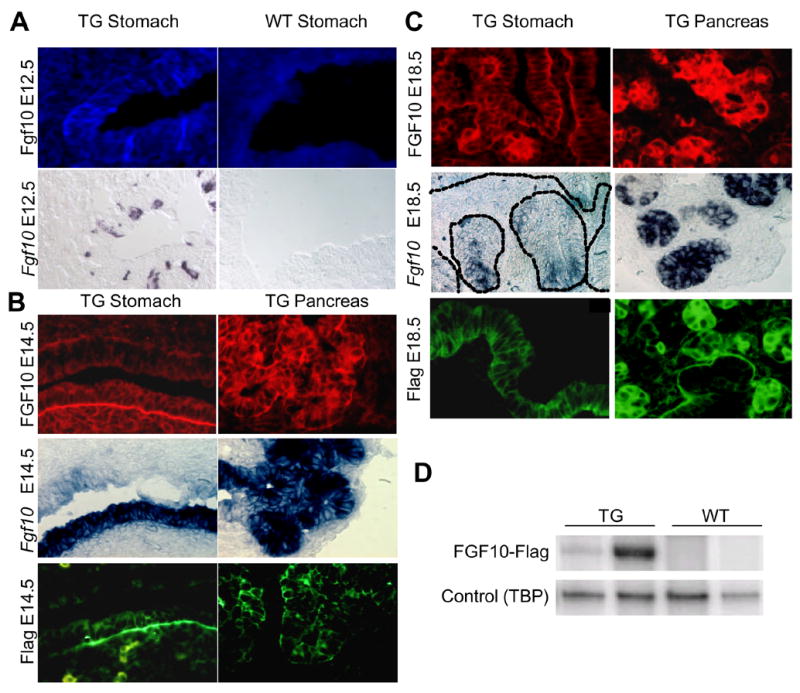
A: E12.5 posterior stomach. Expression of FGF10 was detected with an antibody that detects only high levels of the protein. Fgf10 mRNA was detected by in situ hybridization on an adjacent slide. A few cells in the most posterior stomach of the TG expressed FGF10. No staining was detected in WT littermates. B: E14.5 posterior stomach (lower epithelium). Flag and FGF10 protein detected by immunohistochemistry and Fgf10 mRNA detected by in situ hybridization. No staining was detected in WT littermates (data not shown) n=3. ISH and IHC were performed on consecutive slides. C: E18.5 antrum. Flag and FGF10 protein detected by immunohistochemistry and Fgf10 mRNA detected by in situ hybridization. No staining was detected in WT littermates (data not shown) n=3. Fgf10 mRNA was only detected in glandular structures at this stage although the protein could also be detected in luminal cells. D: Semi-quantitative RT-PCR for the Fgf10-Flag construct in E18.5 stomach of two transgenic mice and their WT littermates. The shown bands are representative examples from three experiments