
Figure 8. Distal foregut patterning.
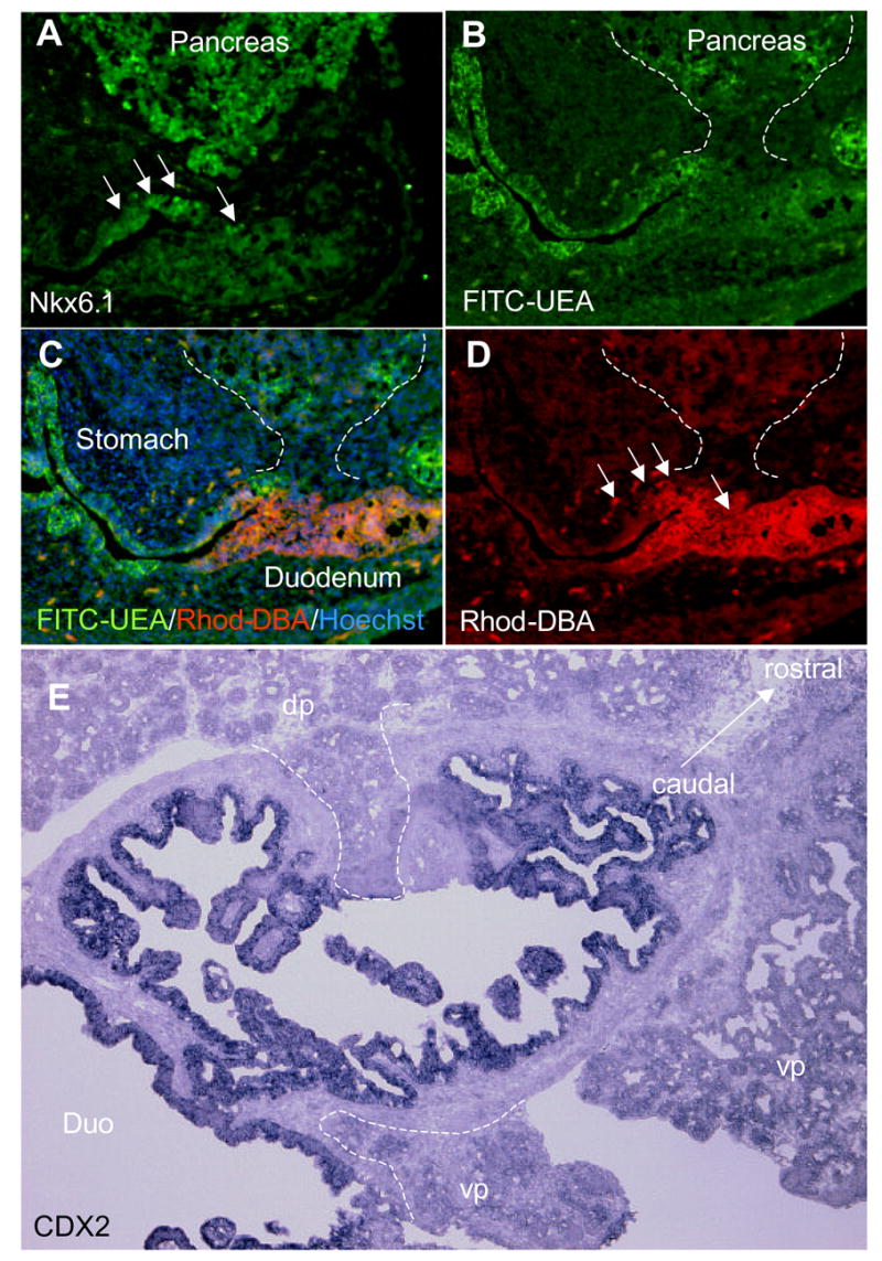
A: Nkx6.1 immunostaining showing the position of the pancreas at E14.5. B: FITC -UEA staining at E14.5. C: FITC-UEA, Rhodamine-DBA and Hoechst staining merge. D: Rhodamine-DBA staining showing the position of the duodenum at E14.5. E: Cdx2 in situ of the duodenum at E18.5