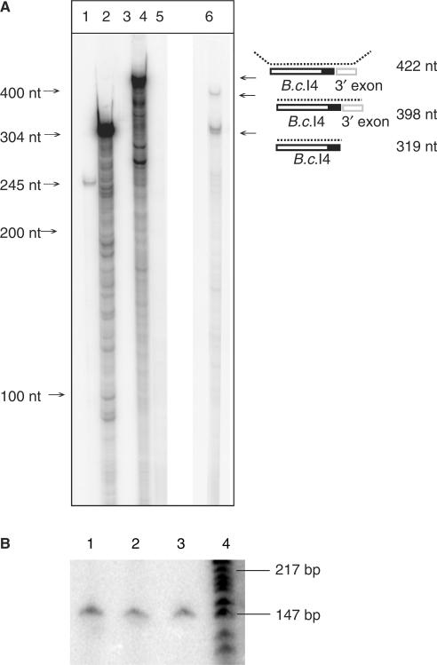
Figure 2.
RNase T1/A protection assay (A) and radioactive RT-PCR (B) showing that the extra 56-nt element 3′ of the B.c.I4 intron is part of the intron RNA and not part of the exons. In A, lanes 1, 2 and 3 show positive controls based on mouse RNA, and lanes 4, 5 and 6 show the results based on B. cereus RNA. Lane 1: digested antisense mouse β-actin RNA probe hybridized with mouse liver RNA; lane 2: same probe as in lane 1, undigested; lane 3: same probe as in lane 1, digested, without mouse liver RNA; lane 4: undigested B.c.I4-3′exon junction probe hybridized to B. cereus ATCC 10987 total RNA; lane 5: same probe as in lane 4, digested, without RNA sample; lane 6: same probe as in lane 4, digested, with RNA sample. A schematic of the experiment illustrating the location of the probe and the expected products is shown on the right. The black area represents the extra 56-nt element. In B, lanes 1, 2 and 3: RT-PCR conducted with exon-specific primers I4B_right (radiolabeled) and I4A_left (Table 1) using as template total RNA sample isolated from B. cereus ATCC 10987 at 3, 4 and 6 h of growth, respectively. Lane 4: γ[32-P]ATP 5′-end-labeled pBR322 DNA digested with MspI (New England Biolabs), as marker.

