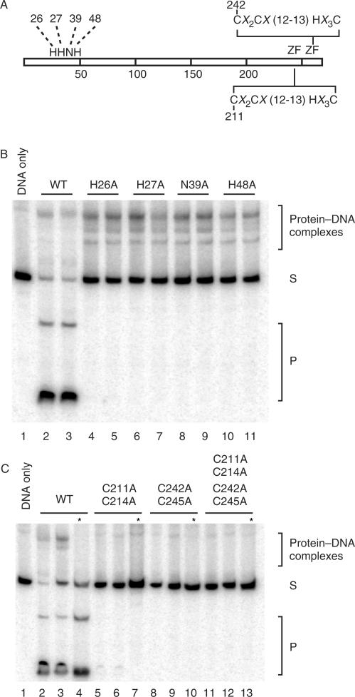
Figure 5.
Cleavage and DNA-binding assays of alanine-substitution mutants. (A) Schematic representation of I-TevIII indicating mutated amino acids. The numbers represent amino acid positions that were changed to alanine. ZF = zinc finger with the consensus sequence. (B) H–N–H mutants. For each mutant assayed, two levels of protein were utilized, 0.2 µg or 0.4 µg. Cleavage and DNA-binding assays are shown. Samples were separated on native 8% polyacrylamide gels. Cleavage products are marked with a P, and unbound DNA with an S. (C) Zinc finger mutants. Analysis and labeling as in panel A. Asterisks represent protease-treated samples.

