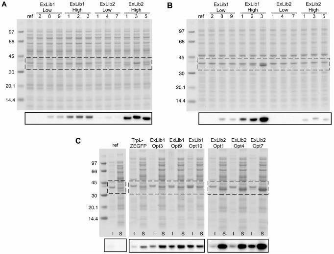
Figure 5.
Analysis of sorted ExLib1 and ExLib2 clones by SDS-PAGE and western blotting. Clones of indicated identities were cultivated and treated as described in the material and methods section for fractionation of soluble and insoluble materials, which were subsequently analyzed by SDS-PAGE under reducing conditions with protein staining or western blotting (smaller insets) using a polyclonal anti-GFP rabbit IgG reagent as primary antibody. The regions of the SDS-PAGE gels corresponding to the western blotting analyses are indicated by the boxes. (A) Soluble fractions from ExLib1-low/high and ExLib2-low/high clones; (B) Insoluble fractions from ExLib1-low/high and ExLib2-low/high clones; (C) Insoluble (I) and soluble (S) fractions from ExLib1-Opt and ExLib2-Opt clones. The lanes designated ‘ref’ refers to samples prepared from plasmid-less host cells (grown without added antibiotic). The numbers indicate molecular weights of reference proteins in kDa (Amersham Biosciences). In (C), samples from the pBR-TrpL-ZEGFP clone was included as additional reference.

