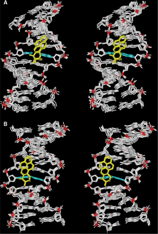
Figure 5.
View into the minor groove of superpositioned intensity-refined structures of the (A) G6*G7 and (B) G6G7* sequence contexts at the 12-mer duplex level. The structures shown are the five best representative conformations for each sequence context from the final 1 ps of unrestrained MD simulation after intensity refinement. The BP rings are in yellow, the modified base guanine and its partner cytosine are in cyan, and the rest of the DNA is in white except for the phosphorus atoms, which are colored red.

