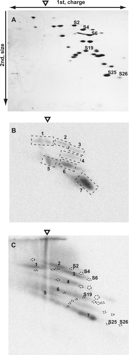
Figure 2.
Identification of a ribosomal protein crosslinked with s4U-labeled PSIV IRES domains 1–2. The origin of the first-dimension gel electrophoresis is indicated by an open triangle. (A) Silver-stained gel after separation of 40S ribosomal proteins. (B) Autoradiograph of a second-dimension gel. The seven signals that were reproducibly detected are indicated by broken squares and numbered from 1 to 7. (C) Autoradiograph of a second-dimension gel containing samples that were incompletely digested with RNase A. The locations of spots of silver-stained proteins are marked with dotted circles. Ribosomal proteins S2, S4, S6, S19, S25 and S26 were identified by mass spectrometry using protein spots excised from a similarly prepared gel.

