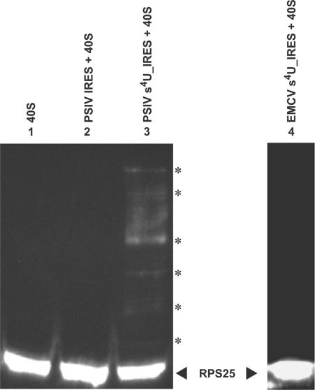
Figure 3.
Evidence of contact between the s4U-labeled IGR-IRES of PSIV and the rpS25 protein of Drosophila. Drosophila 40S ribosomes were mixed with each RNA and irradiated with a near-UV lamp. After partial RNase A digestion, the reaction mixtures were separated on SDS-polyacrylamide (15%) gels and rpS25 proteins were detected by western blot. Lane 1, 40S ribosome with no irradiation; lane 2, 40S ribosome irradiated with unlabeled PSIV IRES; lane 3, 40S ribosome irradiated with s4U-labeled PSIV IRES; lane 4, 40S ribosome irradiated with s4U-labeled EMCV IRES. The position of the rpS25 protein is marked on the right. Asterisks indicate the positions of crosslinked rpS25 proteins in lane 3.

