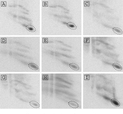
Figure 5.
Identification of uracil residues of the IRES that interact with rpS25. Shown are autoradiographs of second-dimension gels separating the Drosophila 40S ribosomal proteins crosslinked with domain 1–2 mutants of the PSIV IRES containing s4U. Signals corresponding to the rpS25 are circled by broken lines. (A) U6012A+U6015A+U6017A mutation; (B) UU6031AA+UUU6036-8AAA mutation; (C) UU6044-5AA mutation; (D) UU6062-3AA+U6066A mutation; (E) UU6073-4AA mutation; (F) UU6082-3CC mutation; (G) UUU6089-91AAA mutation; (H) UU6096-7CC mutation and (I) U6130A mutation.

