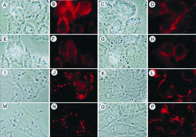
Figure 7.
Effect of CLAMP on the cellular distribution of fluorescent lipid (DiI) delivered to cells from DiI-HDL via SR-BI. CHO cells (A, B, E, F, I, J, M, and N) and CHO-CLAMP cells (C, D, G, H, K, L, O, and P) were plated in a 24-well plate containing a glass coverslip (12 × 12 mm) and transfected with either SR-BI (A–D and I–L) or SR-BIΔC15 (E–H and M–P). The cells were washed once with medium A, and medium B containing 10 μg of protein/ml of DiI-HDL was added for 5 min. The cells were then washed twice with medium A and incubated for 0 h (A–H) or 1 h (I–P) in medium A, after which, the cells were washed with PBS six times followed by fixation in 3.7% (vol/vol) formaldehyde in PBS for 20 min. The cells were examined for the localization of DiI-HDL with a fluorescence microscope (Zeiss).

