
Fig 5. Ax2 binds to Cdc42 at the AP PM.
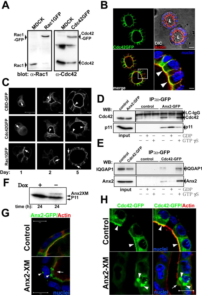
(A) Western blot of stable expression of GFP-Rac1 or GFP-Cdc42. Extracts from MDCK GFP-Cdc42, GFP-Rac1 or control cells were inmunoblotted with anti-Rac1 (left panel) and anti-Cdc42 (right panel) to detect endogenous and transfected proteins.
(B) GFP-Cdc42 distribution in mature cysts. GFP-Cdc42 cells were grown for 5d and stained for nuclei (blue) and actin (red). Bottom-right panel shows the magnification of the indicated region of the merged panel (bottom left). Arrowheads indicate colocalization of GFP-Cdc42 and actin at the AP PM. Scale bar, 10 μm in upper-right panel and 2 μm in the bottom-right panel.
(C) Distribution of CBD-GFP, GFP-Cdc42 and Rac1-GFP during cystogenesis. MDCK CBD-GFP (top row), GFP-Cdc42 (middle row) or GFP-Rac1 (bottom row) cells were grown for 1, 2 or 5d and visualized. Arrows indicate fluorescent proteins at cell-cell or cell-ECM junctions. Arrowheads indicate the localization of CBD-GFP and GFP-Cdc42 at the AP surface. Scale bars are 5 μm or 10 μm as indicated.
(D) Ax2-GFP interacts with endogenous Cdc42 in a GTP-dependent manner. MDCK Ax2-GFP or control cells in mature cysts were lysed and extracts were loaded with GDP or GTPγS. Extracts were immunoprecipitated with antibody against GFP and immunoblotted to analyze Cdc42 and p11. 3% of input or 30% of immunoprecipitated material were loaded on the gel. A band corresponding to the light chain (LC) of IgG was detected with the anti-Cdc42 antibodies and served as a loading control.
(E) Cdc42-GFP interacts with endogenous Ax2 in a GTP dependent manner. Cdc42-GFP or control cells in mature cysts were lysed and the extracts loaded with GDP or GTPγS. Extracts were immunoprecipitated using antibodies against GFP and immunoblotted to analyze Ax2 and IQGAP1. 3% of input and 30% of immunoprecipitated material were loaded in the gel.
(F–H) Effect of Tet-off inducible adenovirus-mediated expression of Ax2X on Ax2-GFP or Cdc42-GFP localization in cysts. MDCK Ax2-GFP (G) or Cdc42-GFP (H) cells formied cysts for 5d were infected with a tet off regulated adenovirus encoding Ax2XM and maintained in the presence (control) or absence (Ax2XM) of 20ng/ml of dox for 16h. Cells were lysed and extracts analyzed by western blot to detect Ax2XM (F). The cysts were and stained for actin (red) and nuclei (blue). Arrowheads indicate aggregates of Ax2-GFP or Cdc42-GFP. Arrows indicate disruption of the AP actin cytoskeleton. Scale bar 10 μm.