
Fig 7. Cdc42 targets aPKC to the AP plasma PM to form the central lumen.
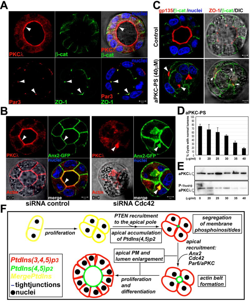
(A) aPKC and Par3 distribute differently in nature cysts. Cysts were stained for aPKCλ (top panels) or Par3 (bottom panels) (red, left panels), β-catenin or ZO-1 (green, middle panels), and for nuclei (merged with DIC, right panels). Arrowheads indicate aPKC at the AP PM, and colocalization of Par3 and ZO-1 at TJ.
(B) Reduction of Cdc42 induces intracellular accumulation of aPKC and Ax2. Ax2-GFP cells were transfected with Cdc42 or control siRNAs and plated to form cysts for 48h. Cells were stained for aPKC (red) and merged with Ax2-GFP and nuclei (bottom-right panels); and actin (red, left-bottom panel merged with DIC). Arrowheads indicate aPKC at the AP PM in controls cells (left panels), or to the intracellular vesicles in Cdc42 depleted cells (right panels).
(C) aPKC-PS disrupts lumen formation. MDCK cysts were treated with aPKC-PS (40μM) or not (control). Cells were stained to detect gp135 (red), β-catenin (green) and nuclei in left panels; or ZO-1 (red), β-catenin (green) and merged with DIC in right panels.
(D) The aPKC inhibitor aPKC-PS disrupts lumen formation in a dose-dependent manner. MDCK cysts were treated with indicated concentrations of aPKC-PS for 48h. Cells were then fixed, stained and quantified for lumen formation. Values shown are mean ±SD from 3 different experiments. N=100/experiment.
(E) aPKC-PS inhibits aPKC phosphorylation in a dose-dependent manner.
(F) Model: PtdIns(3,4,5)p3 (red) and PtdIns(4,5)p2 (green) colocalize in unpolarized MDCK cells (yellow). AP recruitment of PTEN induces the accumulation of PtdIns(4,5)p2 at the AP domain. PtdIns(4,5)p2 recruits Ax2, Cdc42 and Par6/aPKC to form the AP PM and lumen.