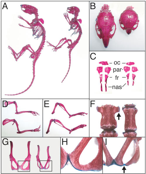
Figure 3. Skeletal defects in R26floxneoWnt4; Col2a1-Cre mutants.
Skeleton preparations from 6-week-old R26floxneoWnt4; Col2a1-Cre mutants and R26floxneoWnt4controls. A, Intact skeletons, control (left) and mutant (right). B, Dorsal view of skulls, control (left) and mutant (right). C, individual dorsal skull bones, control (left) and mutant (right). D, Isolated forelimbs, mutant (top) and control (bottom). E, Isolated hindlimbs, mutant (top) and control (bottom). F, Third lumbar vertebrae, showing lateral ossifications (arrow) in the control (left) that are hypoplastic in the mutant (right). G, Isolated pelvic bones from 6-week-old mice, control (left) and mutant (right). H, Higher magnification of boxed region shown in panel G, showing pubic and ischial bone fusion of the control. I, Higher magnification of boxed region shown in panel G, showing lack of fusion between the pubic and ischial bones of the mutant. fr, frontal bone; nas, nasal bone; par, parietal bone; oc, occipital bone.

