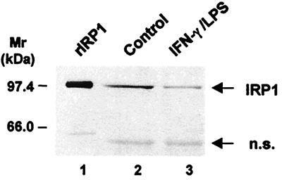
Figure 1.
Regulation of IRP1 expression in RAW 264.7 macrophages upon exposure to a combination of IFN-γ and LPS. RAW 264.7 cells were stimulated with 10 units/ml IFN-γ and 50 ng/ml LPS for 16 h. IRP1 levels in cytosolic extracts were analyzed by Western blotting with an affinity-purified rabbit IRP1 antiserum, as described in Materials and Methods. Purified recombinant IRP1 (rIRP1) was used as a positive control. Molecular masses of protein standards in kDa are shown on the left. The additional band at <66 kDa detected in some experiments (nonspecific, n.s.) may represent cross-reactivity with other protein or a degradation product, as previously observed (24).

