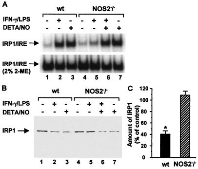
Figure 4.
IRP1 levels in stimulated peritoneal macrophages from wild-type and NOS2 knockout mice. Peritoneal macrophages from wild-type (wt) and NOS2 knockout (NOS2−/−) mice were activated with a combination of 10 units/ml IFN-γ and 50 ng/ml LPS, and/or exposed to 500 μM DETA/NO for 16 h. (A) IRP1–IRE binding activity in cytosolic extracts was analyzed by EMSA. (B) IRP1 levels were analyzed by Western blotting using anti-IRP1 antiserum. (C) IRP1 levels in stimulated cells were quantified by densitometry and are expressed as percentages of controls. Results from three independent experiments are shown. *, Significantly different relative to control levels, P < 0.007.

