
Fig. 3.
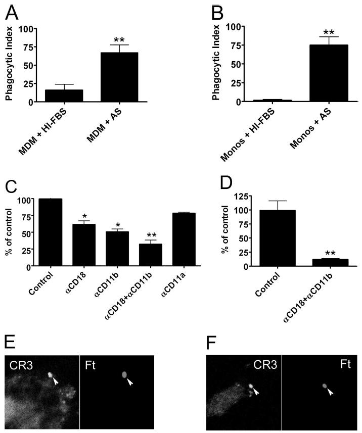
Serum complement is required for optimal phagocytosis of LVS. (A and B) Ft LVS was added at a MOI of 20:1 to MDM (A) or monocytes (Monos; B) in RPMI supplemented with 10% HI-FBS or 2.5% fresh AS as indicated. Phagocytosis was quantified after 1 h at 37°C. Data are the average ± SEM from three to six independent experiments performed in triplicate. **, P < 0.01. (C) MDM in RPMI containing 2.5% AS were left untreated (Control) or incubated with 25 μg/ml anti-CD18, anti-CD11b, anti-CD11a, or a mixture of anti-CD18 and anti-CD11b blocking antibody prior to addition of Ft. Phagocytosis was quantified after 1 h at 37°C. Data are the mean ± SEM from three independent experiments performed in triplicate and are normalized to the no-antibody control (42±7 Ft/100 MDM). *, P < 0.02; **, P < 0.01. (D) Effect of 25 μg/ml anti-CD18 and anti-CD11b antibody on Ft infection of freshly isolated blood monocytes in RPMI containing 2.5% AS. Data are the mean ± SEM from three independent experiments performed in triplicate and are normalized to the no-antibody control (44±4 Ft/100 monocytes). **, P < 0.01. (E and F) Human MDM (E) and monocytes (F) were infected with LVS in RPMI + 2.5% AS for 5 min. Confocal sections show enrichment of CD11b on forming phagosomes (arrowheads).