
Figure 1.
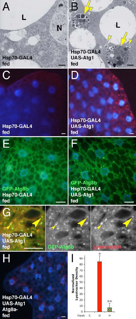
Overexpression of Atg1 induces autophagy.
(A-G) Induction of autophagy by heat shock-induced overexpression of Atg1. TEM showing no induction of autophagy in the fat body of normally fed animals by heat shock alone (A), whereas heat shock-induced overexpression of Atg1 (B) results in the accumulation of autolysosomes (arrows) and autophagosomes (arrowheads). Heat shock alone shows no Lysotracker staining (C), while heat shock-induced Atg1 overexpression results in punctate Lysotracker staining (red) (D). Induction of Atg1 overexpression leads to a redistribution of GFP-Atg8b (green) from uniform staining (E) to a punctate pattern (F). Many GFP-Atg8 positive punctae are found either adjacent to (small arrowheads) or overlapping with (large arrowheads) Lysotracker labeled structures (G). N indicates the nucleus in (A); L indicates a lipid droplet in (A & B).
(H) Mutation of Atg8a disrupts Atg1-induced Lysotracker staining.
(I) Quantitation of Lysotracker staining. * indicates a significant difference from wild type: p < 0.0001; ** indicates a significant difference from Atg1 overexpression alone: p = 0.0001. Error bars indicate standard deviation (SD).
Scale bars represent 1 μm in (A-B) and 10 μm in (C-H). Nuclei are marked in blue in panels (C-H). Genotypes: (A, C) Hsp70-GAL4/+. (B, D) Hsp70-GAL4/UAS-Atg19. (E) Hsp70-GFP-Atg8b6B/Hsp70-GFP-Atg8b6B; Hsp70-GAL4/TM6B. (F-G) Hsp70-GFP-Atg8b6B/Hsp70-GFP-Atg8b6B; Hsp70-GAL4/UASAtg16B. (H) Atg8aKG07569/Y; Hsp70-GAL4/UAS-Atg16B. (I) As in panels C, D, & H.