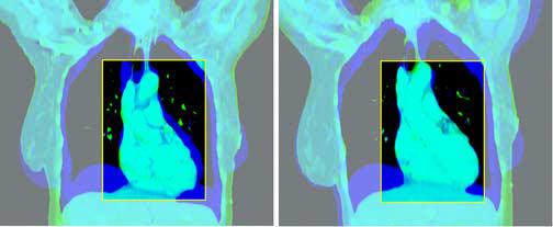
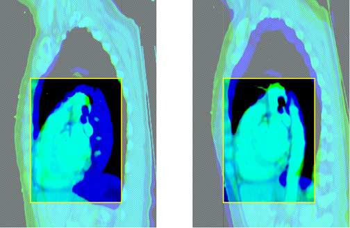
Figure 3.


Rigid Regional Alignment (this figure is in 2 2-part panels, each in its own file: the first panel should be subtitled “Coronal”, with the first part labelled “Before” and the second part labelled “After”; the second panel should be subtitled “Sagittal”, with the first part labelled “Before” and the second part “After”).
This figure depicts the outcome of the rigid regional alignment process. Here, a patient's scan at 80% of vital capacity was aligned to her scan in the exhale state from the same scan session.
