
Figure 7.
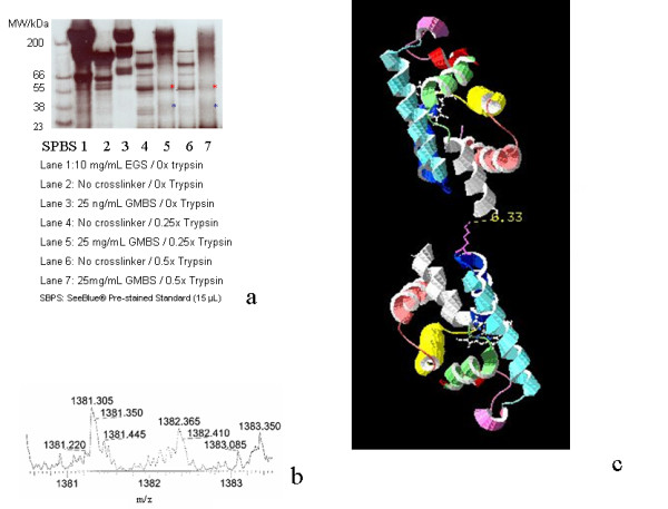
(a) Partial trypsinolysis of EGS-crosslinked HbII using 1.5-hour incubation time, 30 μL of samples were loaded on each lane unless otherwise mentioned. (b) The only genuine crosslinked peaks, the 1381.3051+ peak, as zoom scan. (c) Orientation of Domain T1 and C1 which allows C25CP and K106TP to be positioned in a distance comparable to the expected 6.8Å (maximum GMBS crossbridge span).