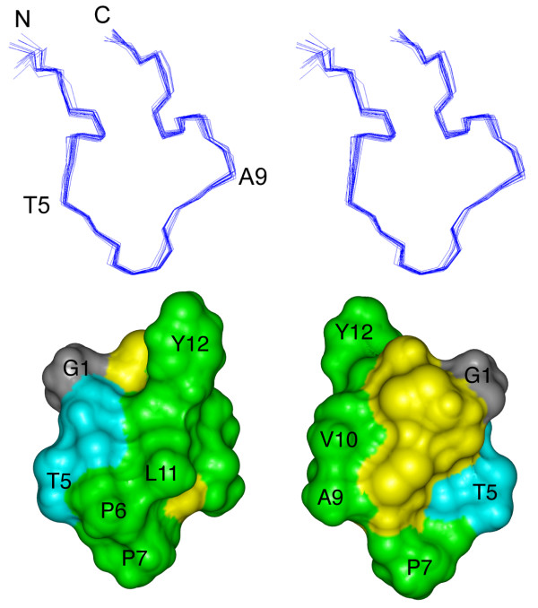
Figure 3.

Three-dimensional structure of the ribbon isomer of BuIA. (top) Stereoview of the 20 NMR-derived lowest energy structures of ribbon BuIA. For clarity only the backbone (N, Cα, C') is shown and the two disulfide bonds are omitted. The structures are superimposed over the whole molecule. N refers to the N-terminus; C refers to the C-terminus. Residues were labeled with the one letter code. (bottom) Solvent accessible surface of ribbon BuIA. The two views are rotated by 180° about the vertical axis. Hydrophobic residues (Pro, Ala, Val, Leu, Tyr) are green, hydrophilic residues (Ser, Thr) are blue, Gly is grey and the Cys residues are yellow. Selected residues are labeled with the one letter code.
