
Figure 3.
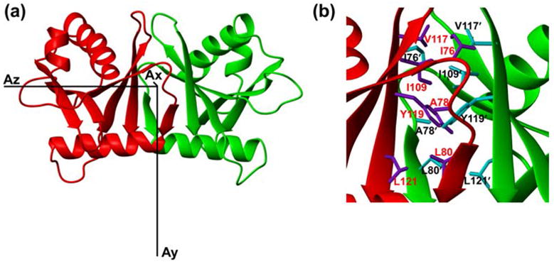
(a) Ribbon diagram of the HsSen15(36–157) structure oriented with regard to three principal axes of alignment tensor. (b) The dimer interfaces of HsSen15(36–157): the side chains of selected residues from one monomer are colored in purple and labeled in red, and those from the symmetric subunit are colored in cyan and labeled in black. Otherwise the color schemes are the same as in Figure 2.