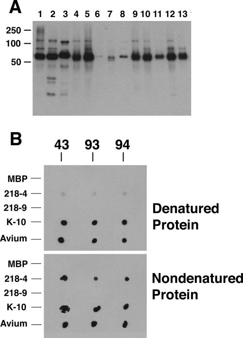
FIG. 6.
Immunoblot (A) and dot blot (B) analyses of aptamers to MAP0105c. (A) The immunoblot containing mycobacterial whole-cell sonicated extracts was exposed to aptamer 94. Lane assignments: 1, M. silvaticum; 2, M. scrofulaceum; 3, M. abscessus; 4, M. avium subsp. paratuberculosis K-10; 5, M. avium subsp. avium (strain TMC702); 6, M. bovis; 7, M. phlei; 8, M. bovis BCG; 9, M. avium subsp. paratuberculosis ATCC 19698; 10, M. avium subsp. avium (strain TMC715); 11, M. avium subsp. paratuberculosis (strain Linda); 12, M. intracellulare; 13, M. kansasii. Size standards are indicated in kilodaltons in the left margin. (B) The dot blot was exposed to the three aptamers, which are indicated above the blots. Proteins spotted to the membrane are indicated in the left margin, and the state of the proteins is indicated in the right margin. Abbreviations: MBP, MBP fused to the α-peptide of LacZ; 218-4, an MBP fusion containing the N-terminal half of MAP0105c; 218-9, an MBP fusion containing the C-terminal half of MAP005c; K-10, a whole-cell lysate of M. avium subsp. paratuberculosis K-10; Avium, a whole-cell lysate of M. avium subsp. avium TMC715.

