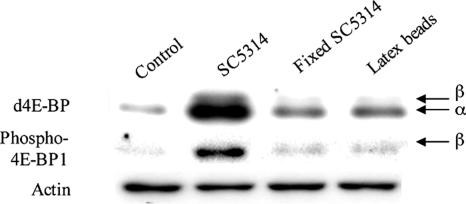
FIG. 4.
Western blot of d4E-BP protein in S2 cells. Lanes from left to right: S2 cells alone (control), S2 cells infected with live C. albicans (SC5314), S2 cells in the presence of paraformaldehyde-fixed C. albicans, and S2 cells ingesting latex beads for 6 h. Identical amounts of total protein (30 μg) were analyzed by Western blotting with 1868 antibody to d4E-BP or phospho-4E-BP1 (thr37/46). α, active, nonphosphorylated isoform; β, hyperphosphorylated isoform; actin, loading control.

